
Introduction
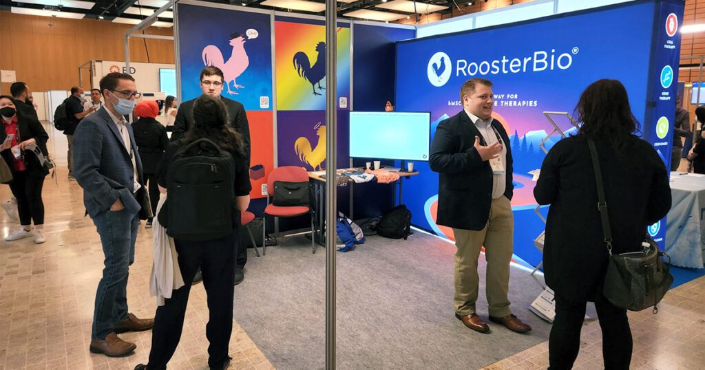
After two years of virtual-only meetings, ISEV 2022 kicked off in-person in the beautiful city of Lyon, France from May 25-29 with an estimated over 1500 attendees, including five from RoosterBio. Six plenary talks from renowned speakers such as Benedetta Bussolati (University of Turin) and Molly Stevens (Imperial College London) served as the nightcaps to active days comprised of over thirty total talk sessions occurring concurrently over the conference period.
Over 400 posters filled the ISEV exhibit hall with booths from companies including NanoFCM, Particle Metrix, and EVerZom. Our team estimated that >50% of booths were associated with analytical tools, with the remainder focusing on research tools, purification, and cell/media supply.
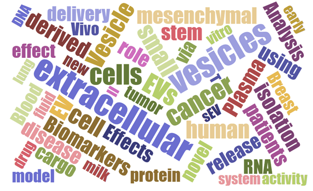
A growing theme at ISEV in recent years is the clinical translation of therapeutic extracellular vesicles (EVs), peaking with each subsequent conference and again at ISEV 2022. This trend is most evident in a partnership between ISEV and ISCT beginning in 2020 to “facilitate collaborations in the areas of unproven cell & gene therapies as well as translation of promising EV research to the clinic” [1]. MSC-EVs are steadily gaining traction as therapeutics, and the word ‘mesenchymal’ is represented in the top 50 words present in ISEV 2022 abstracts (Figure 2).
For the purposes of this blog, we use the nomenclature “EVs” meaning “extracellular vesicles”, which includes the EV subpopulation “exosomes” that is sometimes used in place of “EVs” by clinical or commercial investigators.
Education Day Highlights
All Rooster attendees joined the ISEV Education Day on May 25, which highlighted the basic knowns and “known unknowns” around extracellular vesicles. Biogenesis, Purification, Content, and Analytics were major topics during the session and, thus, were chosen for elaboration below.
Biogenesis
Clotilde Théry (Curie Institute) detailed the current understanding of EV biogenesis and secretion, with a recent publication from their group showing that over short time periods, ectosomes contain primarily CD9 and CD81 but exosomes can contain CD63, CD9 and CD81. This means that exosomes are difficult to distinguish from the broader EV population. Dr. Théry concluded, “There are no exclusive markers of exosomes versus ectosomes validated in all possible cell models,” with an eye on the identification of new markers such as LAMP1, TSPAN4, among others [2].
This talk set the tone for EV heterogeneity as a prominent theme at this meeting, with several prominent EV researchers commenting that EVs comprise an elaborate mixture that is found to be more and more heterogeneous since the inception of ISEV in 2012 (admittedly, heterogeneity is a perennial major theme at ISEV meetings). Efforts are still underway to dissect the heterogeneity, such as a recent paper from Raghu Kalluri’s team (MD Anderson Cancer Center) demonstrating that syntenin-1 could represent a universal EV biomarker [3].
Purification
When considering purification of any biologics, an intrinsic tradeoff exists between purity and yield. Given the heterogeneity of EV populations described above, this inverse relationship becomes ever more important, and it means that “every step of EV isolation can affect the EV composition”, according to Metka Lenassi (University of Ljubljana, Slovenia). Throughout ISEV, it became clear that small-scale isolation procedures typically involve ultracentrifugation and/or size exclusion chromatography (SEC), while larger-scale preparations prefer tangential flow filtration (TFF) and/or bind-elute chromatography.
Content
Once EVs are purified, what contents within EVs are the most important, and how can we measure the presence and amount of that content? RoosterBio is intensely focused on this question, and this question was repeated often at ISEV 2022. As a surprise to some unfamiliar with the associated research, Esther Nolte-‘t Hoen (Utrecht University) revealed the detection of Y-RNAs and tRNAs in EVs, while reaffirming the presence of well-known nucleic acid contents miRNAs and mRNAs.
A debate ensued about whether all cargoes within EVs are necessarily relevant from a functional perspective. The debate centers around a recent publication in PLOS Genetics entitled “MicroRNAs are minor constituents of extracellular vesicles that are rarely delivered to target cells”, which claims that although present in EVs, miRNAs are rarely functionally delivered EV cargos without the expression glycoprotein VSV-G on the EV surface [4].
Many key opinion leaders weighed in on the debate throughout the ISEV Education Day. Bernd Giebel (University Hospital Essen) added that one proposed function of extracellular vesicles is “taking out the trash”, or disposing of unwanted cellular contents, and therfore some components within EVs may simply be waste material – a material that may or may not be functionally relevant. Kenneth Witwer (Johns Hopkins) added the broader point that components within the secretome can all be considered relevant depending on a context of utility – for diagnostic biomarkers, or therapeutics, whether materials are encapsulated in exosomes, apoptotic bodies or exomeres, etc., and thus it is useful to consider a broader understanding of EVs in relationship to the entire secretome.
Analytics
To tackle the questions of heterogeneity and content, the most prominent trend of ISEV 2022 was single-EV analysis. “Single-vesicle platforms can address EV intrinsic diversity and heterogeneity”, said Marina Cretich (SCITEC, Milan), which includes tools such as nanoparticle tracking analysis, nano-flow cytometry, super-resolution microscopy, and Raman tweezers microspectroscopy.
Nano-flow cytometry is becoming a widely used technique, with a handful of companies attending ISEV 2022 to demonstrate their technology. As such, Joanne Lannigan (Flow Cytometry Support Services) detailed the DO’s and DON’T’s of EV flow cytometry. In summary, assumptions made for cell-based flow cytometry do not hold for EV-based flow cytometry and thus, EV protocols need to be developed from scratch with careful control. A given EV surface likely holds an estimated 1000- to 100,000-fold fewer antigens than a given cell surface, and thus detection can be problematic even for positive vesicles. In addition, proper reference materials with properties more like EVs and not synthetic nanoparticles are needed in this effort.
Nevertheless, advances in single-EV analytics are certainly at their most advanced state, and throughout ISEV it was encouraging to continually see the advances in EV analytics begin to provide clarity and synthesize a foundation of knowledge regarding the EV heterogeneity. Though researchers continue to unearth novel observations pointing to EV heterogeneity, advances in single-EV analytics are working to augment these discoveries.
Session & Poster Highlights: A Deeper Dive into Major ISEV Themes
Heterogeneity
A large portion of ISEV includes the rich and exciting fields of EVs derived from biological fluids such as blood and milk. Those working with cell culture derived EVs stand to benefit from discoveries made in these fields.
- Blood contains a plethora of EVs which are typically studied to discover and utilize biomarkers. It is well-understood that EVs from serum do not resemble those from cell culture, in terms of tetraspanin expression [5]. Additionally, blood contains an abundance of lipoproteins which are particulate but not considered EVs. Jilian Bracht (University of Amsterdam) estimated that lipoproteins outnumber EVs in blood plasma by 5 orders of magnitude (1016/mL versus 1011/mL). Lessons learned from purification of blood plasma derived EVs will be profoundly useful for EV bioprocessing. However, purifying proteins out of blood may be problematic for stability, as Britta Bettin (University of Amsterdam) commented that absence of protein in purified blood EVs caused them to attach to plastic walls after thawing. Thus, the amount of protein within final EV preparations must be considered for optimal formulation stability.
- Milk is another abundant source of EVs, containing over 1011 EVs per mL on average – even after purification by density gradient centrifugation and SEC, as reported by Martijn van Herwijnen (Utrecht University). These EVs can be used to study nutrition or even used as an EV source for applications such as drug delivery. With so many EVs, heterogeneity becomes seriously amplified, especially since the original source of EVs in milk likely varies from different cell types in the body. Martjin showed that pre-treatment of milk before purification, such as by acidification or treatment with EDTA or sodium citrate, is essential for purification process by limiting heterogeneity primarily through removing non-EV particles [6].
Phosphatidylserine (PS) is a well-known phospholipid that is conventionally understood as being displayed on the surface of cells because of apoptosis-dependent “flippase” activity. Originally, it was thought that surface-displayed PS on EVs could be a marker of apoptotic bodies, a subtype of EVs produced during apoptosis. However, it has since been shown that exosomes [7] and ectosomes [8] can be enriched in PS as well, making some to consider PS as a more general EV marker. Researchers at FUJIFILM developed an EV isolation method based on TIM4, a protein that binds to PS. They show that a majority of EVs from different cell types display PS on their surface, supporting the idea that PS could be a broader EV marker. Moving forward, TIM4-based EV isolation of PS+ EVs will be an important tool for more general studies of EV heterogeneity, and potentially also as a method for EV purification.
Colin Hisey (Ohio State) showed a novel method for discriminating the presence of fetal bovine serum (FBS)-derived EVs versus cell-derived EVs using Surface Enhanced Raman Spectroscopy (SERS) combined with machine learning algorithms. Using this method, the percentage of EVs in a sample can be calculated as derived either from FBS or from cell culture. This method will be critical for development of assays to show the presence of contaminating EVs in final EV preparations.
Analytics
Raman spectroscopy in the form of SERS is becoming a popular trend to provide a ‘fingerprint’ for EV composition. Current iterations of this technology perform bulk measurements of total EV populations [9] to obtain ‘fingerprints’ which can be related to purity. However, Molly Stevens and their group developed a single-particle analysis method utilizing Raman technology termed Single Particle Automated Raman Trapping Analysis (SPARTA). This technology uses optical tweezers to trap single nanoparticles and provides the Raman spectrum as a readout. Since the original publication [10], the group has extended the technology to EVs, which can provide a composition analysis of nucleic acids, proteins, and lipids at the single-EV level. This technology will be critical when teasing out the heterogeneity of mixed EV preparations.
Posters shown by Cellarcus, Inc. showcased nano-flow cytometry and demonstrated first that different cell surface cargo molecules are packaged into EVs in different amounts, further solidifying the conference theme of EV heterogeneity, especially in terms of identity markers. The second poster demonstrated that EVs collected from HEK293T cells in a hollow-fiber bioreactor show expression of CD9, CD63, and CD81 as analyzed by flow cytometry, but their expression levels constituted <20% each of the total EV population. Our team estimated that level of expression was consistent across other talks and posters at ISEV 2022, demonstrating the need for more robust analysis of EV identity and a re-evaluation of conventional EV identity markers.
To expand on this, the work presented by Yohan Kim (Mayo Clinic) tackled dim fluorescent signals and reproducible analysis using nano-flow cytometry. This work demonstrated clearly that fluorophores with greater brightness are ideal to show true positive signals, and it may be necessary to use antibodies with a higher degree of fluorophore conjugation to achieve an appropriate brightness. The work suggests that a special grade of antibodies customized for EV-based nano-flow cytometry may be necessary to use versus those for cell-based conventional flow cytometry.
Advances in labeling EVs include the efforts of Beatriz Salinaz (IiSGM, Madrid) and their group to explore (1) radioactive labeling of EVs with 99mTc [11] and (2) covalent labeling of the EV surface through sulfo-cyanine 7.5 [12]. These efforts will provide a much-needed method to label intact EVs without artifact generation. Olesia Gololobova (Johns Hopkins) emphatically echoed this point to the session audience while presenting her work comparing fluorescent membrane dyes using nano-flow cytometry. When testing the dyes MemGlowTM, PKH, and DiO, although each dye also stained the lipoproteins present in plasma samples, each dye showed positive events for EVs, and MemGlowTM showed the greatest consistency across measurements with ~75% total population positivity.
Enrichment of CD73 on MSC-derived EVs was shown previously [13], and many at ISEV presented data showing CD73 as an EV identity marker for MSC-EVs. One function of CD73 is to hydrolyze and convert AMP to adenosine and inorganic phosphate. Measurement of this reaction in a controlled setting can be used as a form of an EV potency assay, shown by some groups including a poster presented by D. Auer (Paracelsus Medical University, Salzburg). Other interesting novel potency assays included a poster presented by Michele Hamrick (Cellular Dynamics) demonstrating cardiomyocyte survival after insult by treatment with EVs. Continual development of novel biological potency assays such as these (and others that might employ off-the-shelf components) will be critical to the translation of EVs as therapeutics.
Therapies
A major theme at ISEV in recent years is the therapeutic effects of EVs, and ISEV 2022 was no different, with at least 6 concurrent sessions dedicated solely to therapeutic applications. The first plenary talk of ISEV 2022 was delivered by Benedetta Bussolati, a true expert in renal pathophysiology and regenerative medicine. The talk reinforced the fact that cellular plasticity is triggered by organ damage, and regeneration proceeds in different ways specific to each organ. Bussolati claimed that MSCs are the prototypical regenerative cell, acting primarily through the secretome, a claim that is reinforced by the great number and vast range of clinical trials involving MSC-EVs.
Though healthy MSCs release EVs, apoptotic MSCs also release EVs, termed conventionally as ‘apoptotic bodies’ or ‘ApoEVs’. It is known that ApoEVs can be therapeutically relevant [14], but whether healthy EVs or ApoEVs are therapeutically relevant material for particular indications remains unclear. To address this, a recent study showed that MSCs rapidly undergo apoptosis after administration in mice, and MSC apoptosis was required for their immunosuppressive efficacy against mouse models of asthma and multiple sclerosis [15]. Further complicating this picture, EVs from healthy versus apoptotic cells are virtually indistinguishable by size or expression of identity markers, though apoptotic MSCs did produce more EVs than healthy MSCs [16]. Future studies will further delineate the identity of ApoEVs versus EVs from healthy cells and their respective roles in treating particular diseases.
EVs with naturally occurring therapeutic potential such as the above are considered native EVs, in contrast to engineered EVs, which are modified to contain therapeutic potential. In engineering EVs for therapies, two broader categories are becoming delineated: (1) endogenous loading through genetic modification of producer cells and (2) exogenous modification of EVs pre- or post-production.
- For (1) genetic modification, multiple groups including Omnia M. Elsharkasy (University Medical Center Utrecht) and Joseph Whitley (University of Georgia) showed the ability to load CRISPR/Cas9 into EVs using genetic modification, but it is yet to be seen whether this will lead to functional consequences in vivo. Wenyi Zheng (Karolinska Institutet, Stockholm) expanded on previous glycan engineering strategies by using genetic engineering to display the glycan sLeX on EV surfaces, leading to greater uptake by endothelial cells.
- For (2) synthetic modification , Nina Erwin (University of Florida) showcased a system to drive packaging of EVs by introducing a nanoparticle-based cellular transfection tool. Once nanoparticles are internalized in cells, a photo trigger release of contents into the endocytic pathway leads to more efficient cargo loading into EVs. Boya Peng (National University of Singapore) demonstrated functional modification of the red blood cell-derived EV surface, expanding this option as a platform.
During ISEV Education Day, EV therapy pioneer Sai Kiang Lim (A*STAR) emphasized that the path to clinical testing is similar for engineered EVs or native EVs. Generally, researchers presenting at ISEV with eyes on clinical translation are working at a smaller scale. To drive home the importance of considering scale-up, Dr. Lim remarked “Until reaching GMP, you cannot appreciate the challenge.” Scalable process parameters are required to produce EV-based drugs that are reproducible in terms of identity and potency. Once there, release parameters must be identified and measured consistently. As the product parameters can change depending on process parameters, including the production scale, it is important to consider scale-up sooner than later to ensure maintenance of critical quality attributes after scale-up.
Raw material inputs including cells and media are key components for all situations where EVs are produced and used for research purposes. Thus, control over these components is massively important for not only maximizing EV productivity and recovery but also the underlying EV biology. RoosterBio is at the forefront of providing raw material inputs, including cells, medium, and EVs, for the development scalable regenerative cures. Thus, attendance at ISEV and involvement with the ISEV community is extremely beneficial to further our collective understanding of MSC-EVs. The RoosterBio team was thrilled to be in attendance for ISEV 2022 and is excited to participate in future developments of best practices for scalability that will be discussed at ISEV in the coming years.
References
- Börger V, Weiss DJ, Anderson JD, Borras FE, Bussolati B, Carter DRF, et al. International Society for Extracellular Vesicles and International Society for Cell and Gene Therapy statement on extracellular vesicles from mesenchymal stromal cells and other cells: considerations for potential therapeutic agents to suppress coronavirus disease-19. Cytotherapy. 2020;22(9):482-5; doi: 1016/j.jcyt.2020.05.002.
- Mathieu M, Nevo N, Jouve M, Valenzuela JI, Maurin M, Verweij FJ, et al. Specificities of exosome versus small ectosome secretion revealed by live intracellular tracking of CD63 and CD9. Nat Commun. 2021;12(1):4389; doi: 1038/s41467-021-24384-2.
- Kugeratski FG, Hodge K, Lilla S, McAndrews KM, Zhou X, Hwang RF, et al. Quantitative proteomics identifies the core proteome of exosomes with syntenin-1 as the highest abundant protein and a putative universal biomarker. Nat Cell Biol. 2021;23(6):631-41; doi: 1038/s41556-021-00693-y.
- Albanese M, Chen YA, Huls C, Gartner K, Tagawa T, Mejias-Perez E, et al. MicroRNAs are minor constituents of extracellular vesicles that are rarely delivered to target cells. PLoS Genet. 2021;17(12):e1009951; doi: 1371/journal.pgen.1009951.
- Mizenko RR, Brostoff T, Rojalin T, Koster HJ, Swindell HS, Leiserowitz GS, et al. Tetraspanins are unevenly distributed across single extracellular vesicles and bias sensitivity to multiplexed cancer biomarkers. J Nanobiotechnology. 2021;19(1):250; doi: 1186/s12951-021-00987-1.
- Kleinjan M, van Herwijnen MJ, Libregts SF, van Neerven RJ, Feitsma AL, Wauben MH. Regular Industrial Processing of Bovine Milk Impacts the Integrity and Molecular Composition of Extracellular Vesicles. J Nutr. 2021;151(6):1416-25; doi: 1093/jn/nxab031.
- Skotland T, Hessvik NP, Sandvig K, Llorente A. Exosomal lipid composition and the role of ether lipids and phosphoinositides in exosome biology. J Lipid Res. 2019;60(1):9-18; doi: 1194/jlr.R084343
- Meldolesi J. Exosomes and Ectosomes in Intercellular Communication. Curr Biol. 2018;28(8):R435-R44; doi: 1016/j.cub.2018.01.059.
- Gualerzi A, Kooijmans SAA, Niada S, Picciolini S, Brini AT, Camussi G, et al. Raman spectroscopy as a quick tool to assess purity of extracellular vesicle preparations and predict their functionality. J Extracell Vesicles. 2019;8(1):1568780; doi: 1080/20013078.2019.1568780.
- Penders J, Pence IJ, Horgan CC, Bergholt MS, Wood CS, Najer A, et al. Single Particle Automated Raman Trapping Analysis. Nat Commun. 2018;9(1):4256; doi: 1038/s41467-018-06397-6.
- Gonzalez MI, Martin-Duque P, Desco M, Salinas B. Radioactive Labeling of Milk-Derived Exosomes with (99m)Tc and In Vivo Tracking by SPECT Imaging. Nanomaterials (Basel). 2020;10(6); doi: 3390/nano10061062.
- Gonzalez MI, Gonzalez-Arjona M, Santos-Coquillat A, Vaquero J, Vazquez-Ogando E, de Molina A, et al. Covalently Labeled Fluorescent Exosomes for In Vitro and In Vivo Applications. 2021;9(1); doi: 10.3390/biomedicines9010081.
- Angioni R, Liboni C, Herkenne S, Sanchez-Rodriguez R, Borile G, Marcuzzi E, et al. CD73(+) extracellular vesicles inhibit angiogenesis through adenosine A2B receptor signalling. J Extracell Vesicles. 2020;9(1):1757900; doi: 1080/20013078.2020.1757900.
- Caruso S, Poon IKH. Apoptotic Cell-Derived Extracellular Vesicles: More Than Just Debris. Front Immunol. 2018;9:1486; doi: 3389/fimmu.2018.01486.
- Pang SHM, D’Rozario J, Mendonca S, Bhuvan T, Payne NL, Zheng D, et al. Mesenchymal stromal cell apoptosis is required for their therapeutic function. Nat Commun. 2021;12(1):6495; doi: 1038/s41467-021-26834-3.
- Skovronova R, Grange C, Dimuccio V, Deregibus MC, Camussi G, Bussolati B. Surface Marker Expression in Small and Medium/Large Mesenchymal Stromal Cell-Derived Extracellular Vesicles in Naive or Apoptotic Condition Using Orthogonal Techniques. 2021;10(11); doi: 10.3390/cells10112948.